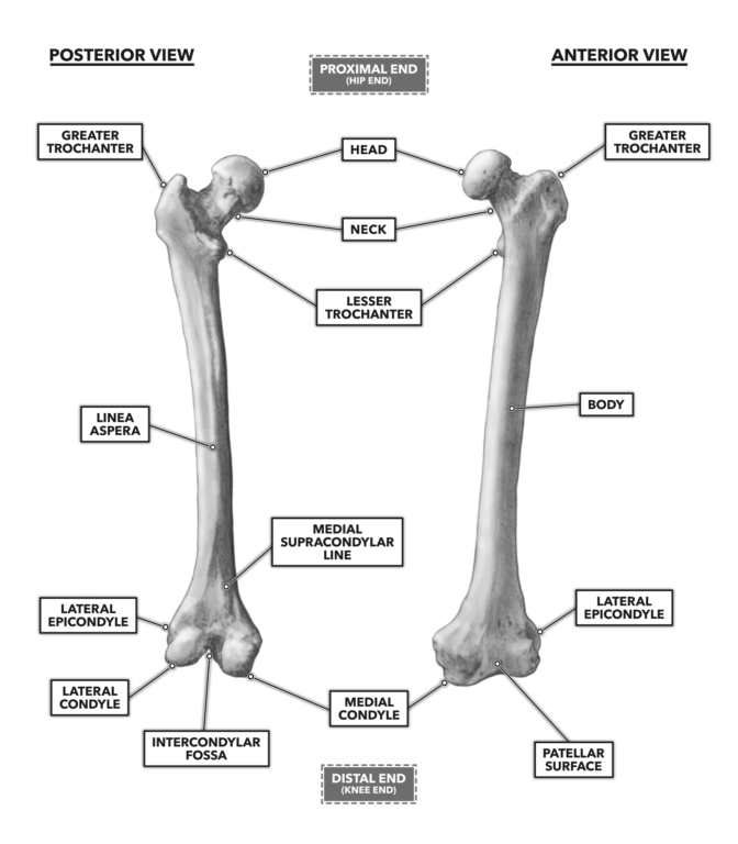
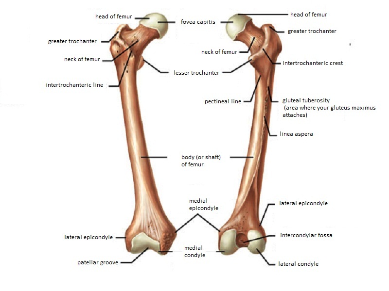
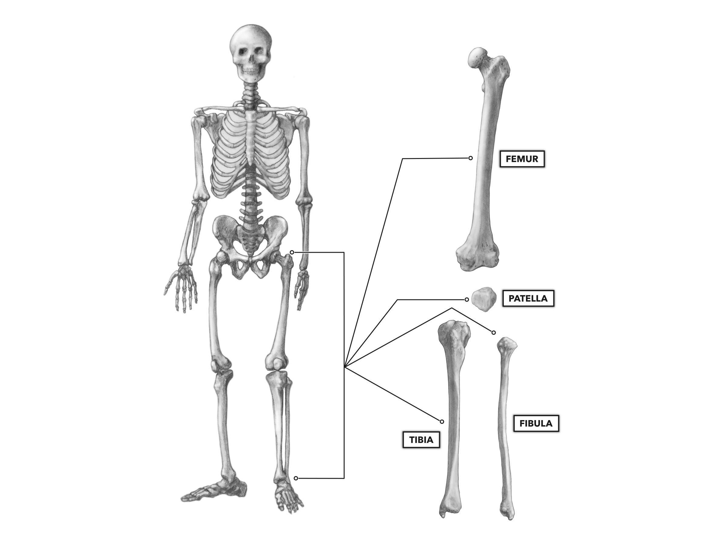
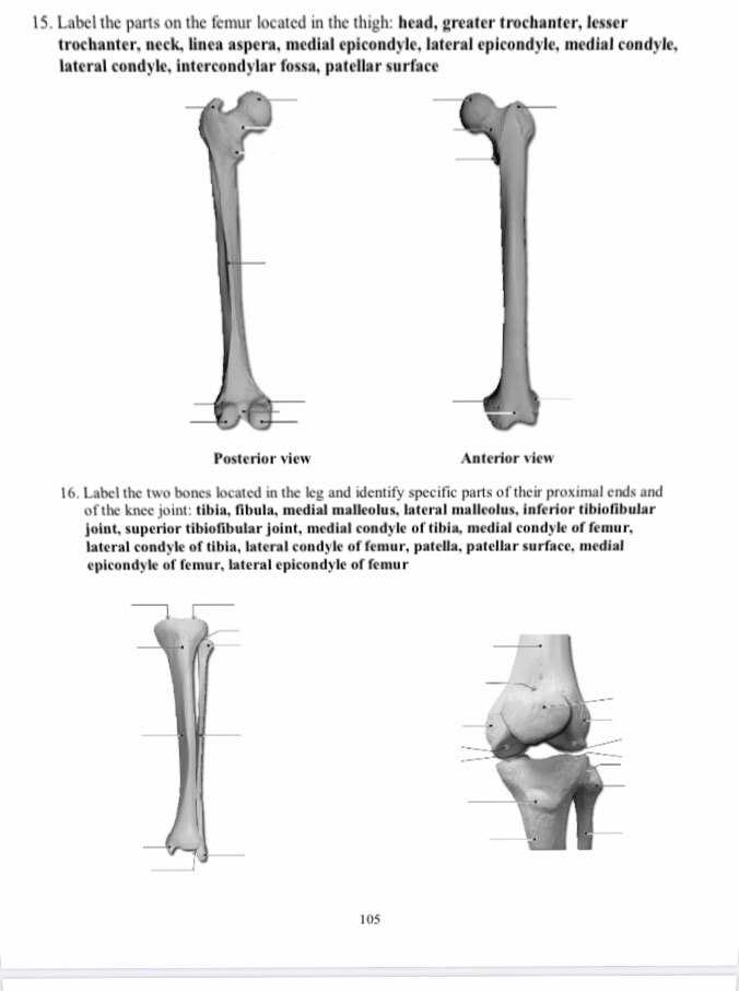
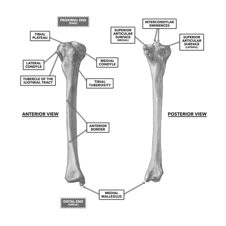
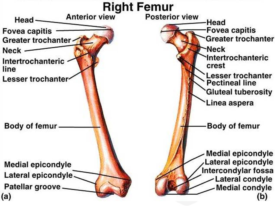
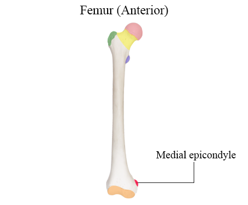
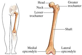
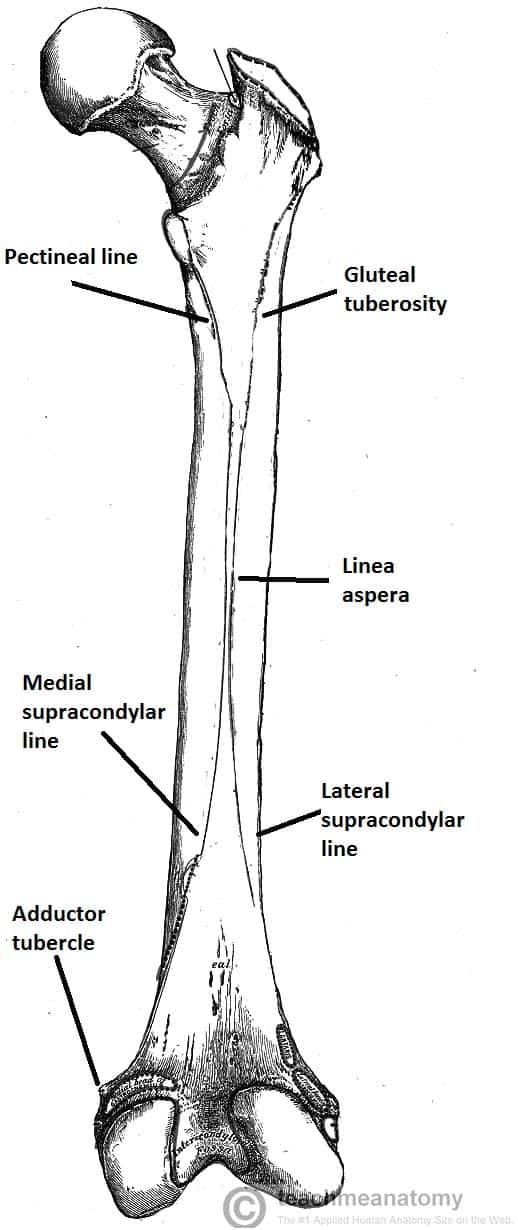
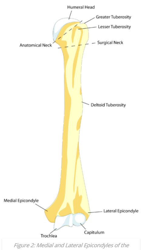
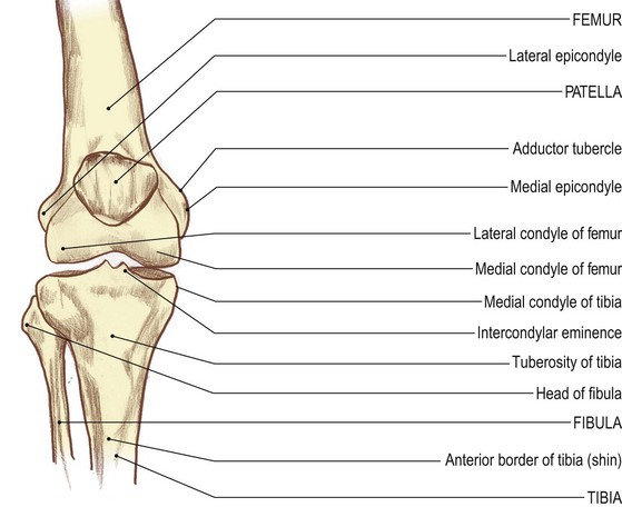
Koleksiyon 2021: femur medial epikondil
Burada aradığınızı bulacaksınız - femur medial epikondil
Ana sayfa
2025 02 15
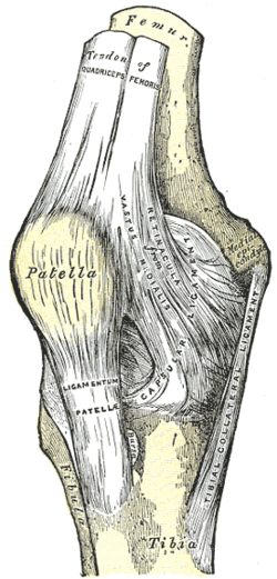
Https www alamy com cunninghams text book of anatomy anatomy vastus media lis ilio psoas lateral epicondyle lateral condyle fig 236the eight femur seen from the front medially and slightly forwards fig 23anterior aspect of proximal por tion of the eight femur with attachments of muscles mapped out bone in the body proximally the femora are separated by the width of the pelvis distally they articulate with the tibiae and patellae in the military position of attention with the knees close to gether the shafts of the thigh bones occupy an oblique position for descriptive purposes the bone is image231857164 html (Dosya tipi jpg)
Cunningham S Text Book Of Anatomy Anatomy Vastus Media Lis Ilio Psoas Lateral Epicondyle Lateral Condyle Fig
Https www alamy com the anatomy of the domestic animals veterinary anatomy trochanteric fossa head y trochanter neck trochanter neck trochanter medial rough line lateral l epicondylc i l medial epicondyle trochlea major nutrient foramen lateral rough line fig 224kight femur op dog anterior view lateral condyle i nlercondyloid fossa fig 225right femur of dog posterior view 1 2 sesamoid bonfts the fibula extends the entire length of the region it is slender somewhat twisted and enlarged at either end the proximal part of the shaft is separated from the tibia by a consider image232326357 html (Dosya tipi jpg)
The Anatomy Of The Domestic Animals Veterinary Anatomy Trochanteric Fossa Head Y Trochanter Neck Trochanter Neck Trochanter Medial Rough Line Lateral L Epicondylc I L Medial Epicondyle Trochlea Major
Https www alamy com anatomy of the woodchuck marmota monax woodchuck mammals 14 10 11 fig 2 38 left femur cranial view and cranial caudal and lateral views of the patella 1 major trochanter 2 neck 3 third trochanter 4 shaft 5 trochlea 6 lateral epicondyle 7 lateral condyle 8 patellar cartilage 9 base of patella 10 articular surface of patella 1 1 apex of patella 12 medial condyle 13 medial epicondyle 14 minor trochanter 15 femoral head formed laterally by the ischium ventrally by the pubis and dorsally by the sacrum and first three to four caudal vertebrae the outlet of the pelvi image236800627 html (Dosya tipi jpg)
Anatomy Of The Woodchuck Marmota Monax Woodchuck Mammals 14 10 11 Fig 2 38 Left Femur Cranial View And Cranial Caudal And Lateral Views Of The Patella 1 Major Trochanter 2 Neck
Https www alamy com anatomy of the woodchuck marmota monax woodchuck mammals 10 11 fig 2 38 left femur cranial view and cranial caudal and lateral views of the patella 1 major trochanter 2 neck 3 third trochanter 4 shaft 5 trochlea 6 lateral epicondyle 7 lateral condyle 8 patellar cartilage 9 base of patella 10 articular surface of patella 1 1 apex of patella 12 medial condyle 13 medial epicondyle 14 minor trochanter 15 femoral head formed laterally by the ischium ventrally by the pubis and dorsally by the sacrum and first three to four caudal vertebrae the outlet of the pelvic can image236800608 html (Dosya tipi jpg)
Anatomy Of The Woodchuck Marmota Monax Woodchuck Mammals 10 11 Fig 2 38 Left Femur Cranial View And Cranial Caudal And Lateral Views Of The Patella 1 Major Trochanter 2 Neck 3
Görüntüler telif hakkına tabi olabilir daha fazla bilgi

